iStar AI Unveils Unprecedented Clarity in Medical Imaging
In a groundbreaking development, researchers at the Perelman School of Medicine at the University of Pennsylvania have introduced an innovative artificial intelligence tool called iStar (Inferring Super-Resolution Tissue Architecture). This tool aims to enhance the interpretation of medical images, providing clinicians with unparalleled clarity and efficiency in diagnosing diseases, particularly cancer.
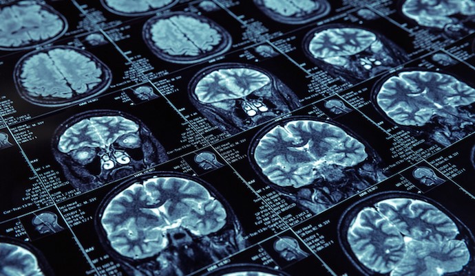
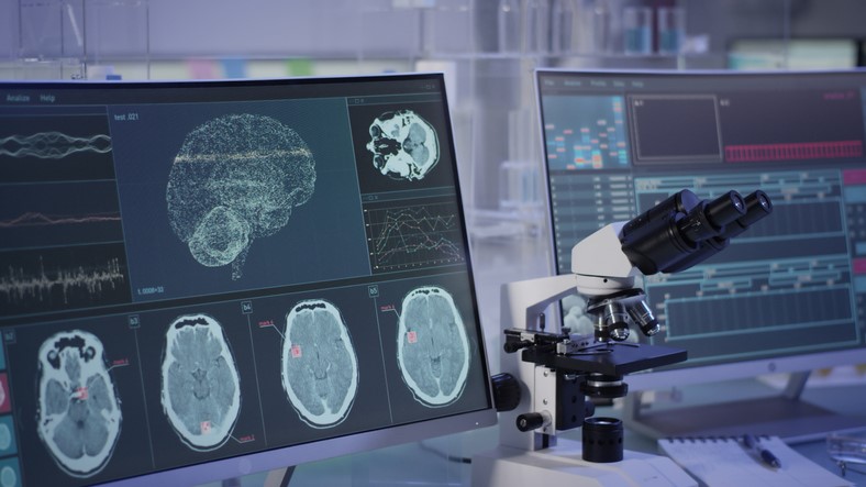
Figure 1. Medical Imaging.
The Breakthrough
Figure 1 is an example of medical imaging. The research, led by Daiwei “David” Zhang, PhD, and Mingyao Li, PhD, introduces iStar as a tool that can potentially transform cancer diagnosis and treatment. By offering highly detailed views of individual cells and a comprehensive look at the spectrum of gene operations, iStar enables the detection of cancer cells that may have gone unnoticed using traditional methods.
Key Features and Applications
iStar employs a unique imaging technique that not only reveals detailed cellular structures but also provides a broader understanding of tissue architecture. This capability is crucial for determining safe margins in cancer surgeries and automating annotations for microscopic images, paving the way for molecular disease diagnosis at an unprecedented level.
Importantly, iStar has the ability to automatically detect critical anti-tumor immune formations known as “tertiary lymphoid structures.” These formations are correlated with a patient’s likely survival and positive response to immunotherapy, making iStar a potential tool for precise patient selection for immunotherapy.
The Technology Behind iStar
Developed as part of spatial transcriptomics, a cutting-edge field mapping gene activities within tissues, iStar utilizes a machine learning tool called the Hierarchical Vision Transformer. This tool breaks down images into different stages, mimicking the process of a pathologist studying a tissue sample. It starts small, identifying fine details, and progressively zooms out to capture broader tissue patterns. The information gathered is then used to predict gene activities, often at near-single-cell resolution.
Clinical Efficacy and Speed
To assess its efficacy, iStar was tested on various cancer tissues, demonstrating its ability to automatically detect hard-to-identify cancer cells. The speed at which iStar operates is a game-changer, completing analyses in just nine minutes compared to competitors that took over 32 hours for similar tasks. This remarkable speed makes iStar applicable to large-scale biomedical studies, 3D analysis, and biobank sample predictions.
Future Directions
As the research team explores further applications, the focus is on extending iStar to 3D contexts and biobanks. The goal is to contribute to a better understanding of microenvironments within tissues, providing valuable data for diagnostic and treatment purposes.
With iStar's revolutionary capabilities, the field of medical imaging is poised for a transformative leap, offering clinicians a powerful tool to enhance cancer diagnosis and treatment planning. As research progresses, the potential applications of iStar in various medical scenarios continue to expand, holding promise for improved patient outcomes and more efficient healthcare practices.
Source: Penn Medicine
Cite this article:
Hana M (2024), China's Breakthrough in Laser Technology: Ultrathin Optical Crystal Revolution, AnaTechMaz, pp. 383
Recent Post
-

The Power of Deep Learning in X-ray Diffraction Analysis for Advanced Materials Characterization
In a groundbreaking study published in npj Computational Materials, scientists...
-
AI-Powered Solution Proposed for Digital Financial Inclusion of Migrant Workers
A proposal put forward by researchers at the Indian Institute of Technology...
-
IIT Madras Develops India-Specific AI Model for Foetal Age Determination
In collaboration with the Translational Health Science and Technology Institute...
-
Accelerating Parkinson’s Drug Discovery with AI
Researchers at the University of Cambridge employed an AI-driven approach...
-
Advancements in AI for Eye Healthcare
A recent study led by the University of Cambridge reveals that GPT-4, a cutting...
-
Advancements in Causal Machine Learning for Medical Applications
An international research team, led by Ludwig-Maximilians-Universität München...
-

The Uncanny Realism of AI-Generated White Faces: A Wake-Up Call for Technology Ethics
Recent research led by experts at The Australian National University...
-
iStar AI Unveils Unprecedented Clarity in Medical Imaging
In a groundbreaking development, researchers at the Perelman School...
-
AI Revolutionizes Heart Health Assessment
In a groundbreaking study, researchers from the Icahn School of Medicine ....
-

Azahar Ali's Vision for the Fourth Agricultural Revolution
As the world braces itself for the fourth agricultural revolution, Azahar...