
Scientists Develop New Method to Freeze and Restore Living Brain Tissue
Scientists have made progress toward safely freezing and restoring brain tissue by preventing the formation of damaging ice crystals, which typically destroy cells during freezing and thawing.
Inspired by the Siberian salamander — an amphibian known for surviving extreme cold — researchers are exploring biological strategies that could one day allow living tissues to be preserved for long periods and later revived without loss of function.
The Siberian salamander is reported to survive extreme cold conditions, including temperatures approaching −50°C and long periods embedded in permafrost, by entering a hibernation-like state and resuming normal activity once temperatures rise.
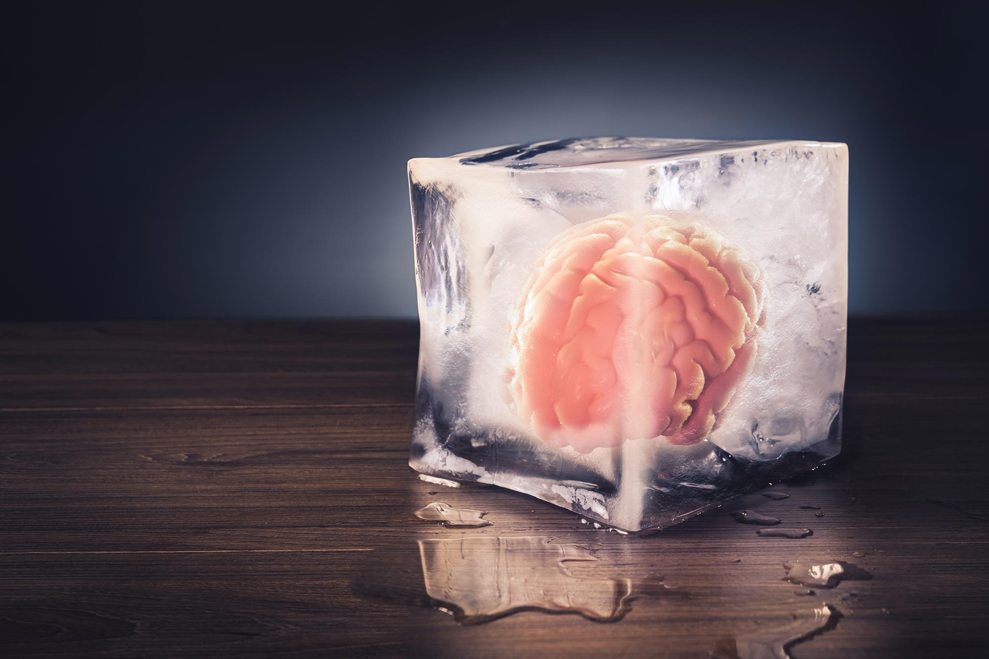
Figure 1. New Cryogenic Methods Aim to Preserve Cells and Neural Networks
Researchers believe this resilience is due to a natural “antifreeze” mechanism. The salamander’s liver produces glycerol, a compound that lowers the freezing point of bodily fluids and helps protect cells and tissues from damage during freezing and thawing. This adaptation prevents the formation of ice crystals, which are typically lethal to most organisms at such low temperatures. Figure 1 New Cryogenic Methods Aim to Preserve Cells and Neural Networks.
Alexander German from the Department of Molecular Neurology at Uniklinikum Erlangen explains that extreme cold is typically damaging to living organisms because ice crystals form within tissues and physically disrupt cellular structures.
He notes that these crystals can mechanically injure cells and destroy the delicate nanostructure of biological tissue, which is why freezing is usually lethal to living systems.
Biological Tissue Fluid Forms a Glassy Solid State
Human embryos can be preserved for long periods using deep-freezing techniques that rely on chemical protectants similar to glycerol, which prevent the formation of damaging ice crystals.
Alexander German explains that when tissue is cooled below −130°C, it does not simply freeze into ice but instead enters a glass-like solid state in which water inside and between cells becomes disordered rather than forming crystals. This process is known as vitrification.
Although vitrification has been successfully used for embryos, it has not yet been achieved for brain tissue in a way that allows full functional recovery after thawing. One major challenge is that antifreeze chemicals can be toxic to sensitive cells. This is particularly problematic for brain tissue, which contains vast networks of interconnected neurons and synapses responsible for communication and function.
Optimized Preservation Methods and Freezing Techniques
Researchers have improved vitrification methods by optimizing both the preservative composition and the cooling process to better preserve neural tissue structure. According to Alexander German, earlier techniques often disrupted synapses and damaged the brain’s delicate network, meaning that even surviving cells could no longer function properly.
In new experiments, the team successfully cooled hippocampal tissue from a rodent brain to −130°C and later analyzed it using electron microscopy. The images showed that the tissue’s nanostructure remained intact after freezing. Once thawed, the hippocampus was able to spontaneously resume normal electrical signaling across neural networks.
Beyond restoring basic activity, researchers including Fang Zheng demonstrated that long-term potentiation — a key mechanism for strengthening synaptic connections — could also be induced after thawing. This process is essential for learning and memory formation, suggesting that the preserved tissue retained not just structure, but functional capability as well.
References:
- https://quantumzeitgeist.com/neutral-atoms-combine-connectivity-scalability/
Cite this article:
Janani R (2026), Scientists Develop New Method to Freeze and Restore Living Brain Tissue, AnaTechMaz, pp.767
Recent Post
-
Scientists Find Common Medications May Change the Gut for Years
A new study suggests that the gut microbiome may preserve ....
-
Scientists Develop New Method to Freeze and Restore Living Brain Tissue
Scientists have made progress toward safely freezing and restoring....
Researchers Compare Multiple Sclerosis Models with Human Tissue to Improve Future Therapies
Researchers have uncovered important differences between two...
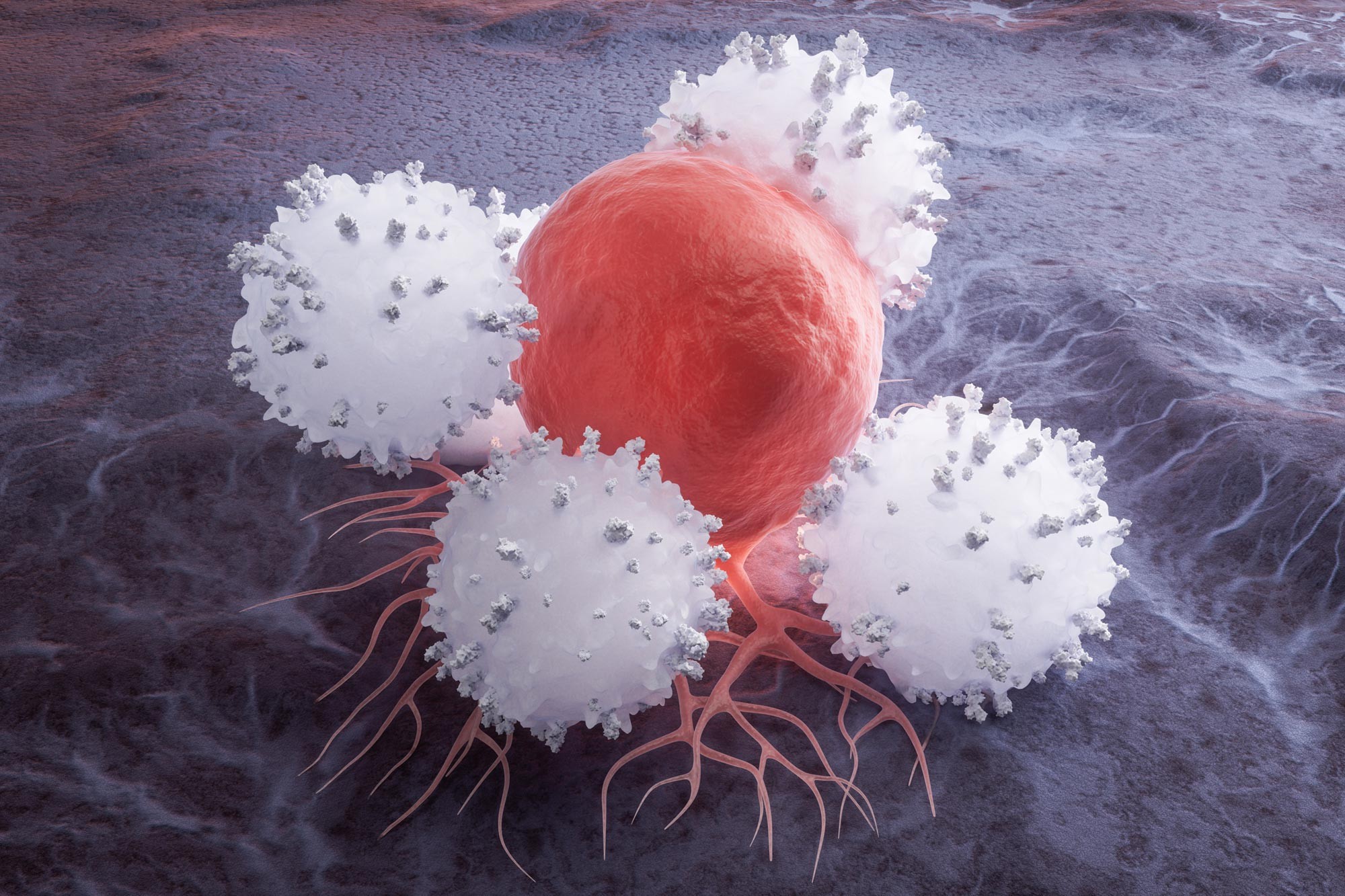
Scientists Identify Genetic “Off Switch” That Boosts CAR T Cell Cancer Fighting Power
A new study has identified a potential strategy for making CAR T-cell....
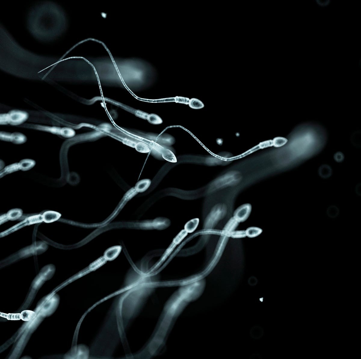
New Research Suggests Sperm Behave Beyond a Fundamental Physics Rule
Sperm cells are able to swim through microscopic environments that....
-
New Strategy Boosts Immune Cells While Starving Tumors
Researchers at UCLA have developed a technique that provides T cells with a protected source of sugar, ....