A Startup Provides Surgeons with A Real-Time View of Breast Cancer During Surgery
MIT spinout Lumicell has developed a drug-device combination that aims to reduce repeat surgeries and improve the completeness of tumor removal. Breast cancer, the second most common cancer and cause of cancer-related deaths among women in the U.S., affects one in eight women. Most women with breast cancer undergo lumpectomy, a procedure to remove the tumor along with a margin of healthy tissue. The excised tissue is then analyzed by a pathologist for any remaining cancer cells at the tissue's edges. However, roughly 20 percent of women who undergo lumpectomies require a second surgery to remove additional tissue.
MIT spinout Lumicell is equipping surgeons with a real-time view of cancerous tissue during surgery, thanks to a handheld device and optical imaging agent that enable the detection of residual cancer cells within the surgical cavity. Surgeons can view these images on a monitor, guiding them to remove any additional tissue as needed during the procedure.
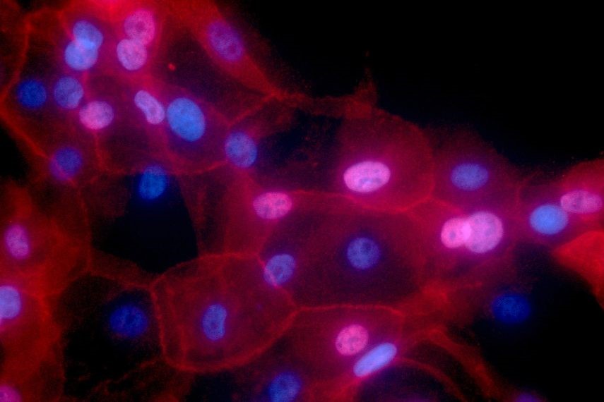
Figure 1. Startup Enables Real-Time Breast Cancer Imaging for Surgeons
In a clinical trial with 357 patients, Lumicell’s technology not only reduced the likelihood of follow-up surgeries but also detected cancerous tissue that could have been missed in a standard lumpectomy. The U.S. Food and Drug Administration approved this technology earlier this year, marking a significant milestone for Lumicell and its founders, MIT professors Linda Griffith and Moungi Bawendi, alongside PhD candidate W. David Lee. Since 2008, much of the system’s development and testing has taken place at MIT’s Koch Institute for Integrative Cancer Research. Figure 1 shows Startup Enables Real-Time Breast Cancer Imaging for Surgeons.
This FDA approval carries a personal significance for some team members, including Griffith, a breast cancer survivor, and Lee, who shifted his career path after losing his wife to the disease in 2003.
A Multidisciplinary Approach
Lee spent 25 years running a technology consulting group before his wife’s breast cancer diagnosis, which inspired him to create technologies that could aid cancer patients. His neighbor, Tyler Jacks, founding director of the Koch Institute, invited Lee to join meetings with professors Robert Langer and Moungi Bawendi at the Koch, and in 2008, Lee became an integrative program officer there. He began working on a new imaging approach using charge-coupled device (CCD) cameras to capture single-cell resolution in living organisms by stabilizing the camera to align CCD pixels with cells, despite movement from an animal’s breathing.
This initiative led to regular meetings with a multidisciplinary group of researchers, including Lumicell co-founders Bawendi (now the Lester Wolfe Professor of Chemistry and 2023 Nobel laureate), Linda Griffith (School of Engineering Professor of Teaching Innovation in Biological Engineering and a Koch Institute faculty member), Ralph Weissleder from Harvard Medical School, and David Kirsch, formerly a postdoc at the Koch and now a scientist at the Princess Margaret Cancer Center. Griffith recalls how these gatherings became collaborative learning sessions: “On Friday afternoons, we’d get together, and Moungi would teach us some chemistry, Lee would teach us some engineering, and David Kirsch would teach some biology.”
During these meetings, they explored the potential of combining Lee’s imaging technique with engineered proteins to illuminate tumor edges where the immune system interacts, useful during surgery. This research received initial funding from the Koch Institute Frontier Research Program through the Kathy and Curt Marble Cancer Research Fund, which Lee describes as critical to their progress: “The Koch Institute provided education, funding, and most importantly, connections to faculty who were willing to teach me biology.”
In 2010, Griffith’s breast cancer diagnosis underscored the importance of their work. “Having that experience as a patient—waiting to hear if my tumor margins were clear after surgery—made me deeply aware of our mission,” Griffith says.
Today, Lumicell’s approach starts two to six hours before surgery, with patients receiving an optical imaging agent through an IV. During surgery, surgeons use Lumicell’s handheld device to scan the cavity walls, displaying any regions likely containing residual cancer on a monitor, which they can then excise. This adds only about seven minutes to the procedure.
Lee highlights the technology’s impact: “It allows surgeons to scan the actual cavity, while pathology only examines the removed tissue, assessing just 1 or 2 percent of its surface. Our system detects residual cancer, reducing second surgeries, and finds cancer in some patients that pathology might miss, which could avoid a second surgery entirely.”
Investigating Additional Cancer Types
Lumicell is currently investigating whether its imaging agent can be activated in other tumor types, including prostate, sarcoma, esophageal, and gastric cancers, among others.
Lee led Lumicell from 2008 until 2020. After stepping down as CEO, he returned to MIT to pursue a PhD in neuroscience—50 years after earning his master’s degree. Howard Hechler subsequently took on the role of Lumicell’s president and chief operating officer.
Reflecting on Lumicell’s journey, Griffith credits MIT’s culture of learning as essential to the company’s creation. “People like David [Lee] and Moungi are committed to solving problems,” Griffith says. “They’re technically brilliant but also love learning from others, which is what makes MIT unique. People are confident in their knowledge yet comfortable not knowing everything, fostering a culture of collaboration. We work together so that the whole is greater than the sum of its parts.”
Source: MIT News
Cite this article:
Janani R (2024), A Startup Provides Surgeons with A Real-Time View of Breast Cancer During Surgery, AnaTechMaz,pp. 265


