MRI Method Detects Luminescent Molecules Deep Within the Brain
A novel magnetic resonance imaging (MRI) technique maps the positions of cells marked with light-emitting molecules even in deep organs and tissues. This method detects changes in blood vessels triggered by bioluminescent proteins, overcoming a major limitation of optical imaging. It holds promise for biomedical applications such as monitoring tumor growth, tracking changes in gene expression, and investigating brain cell activity.
Biologists commonly use light-emitting proteins to label cells, facilitating the study of cellular processes like signaling and metabolism. However, these proteins, effective for intracellular studies, are less suitable for imaging deep tissue structures due to significant absorption and scattering of visible light.

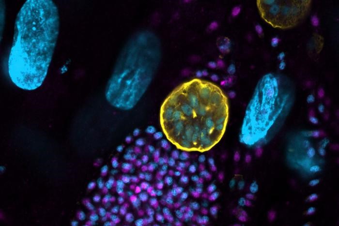
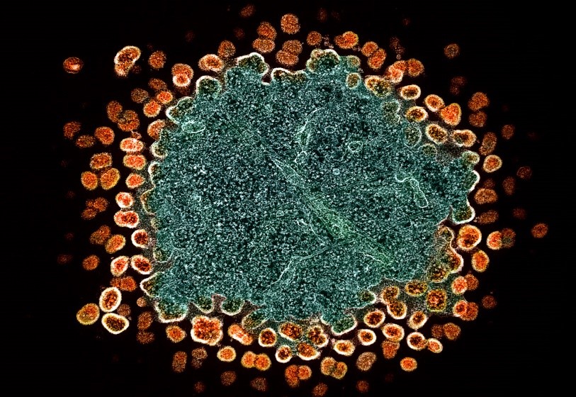
Figure 1. MRI Reveals Luminescent Molecules in Deep Brain Regions
Identifying The Origin of Light Emission
Alan Jasanoff and his team at the Department of Biological Engineering, Massachusetts Institute of Technology (MIT), have developed a novel approach to detect bioluminescence. Their method involves genetically modifying blood vessels to contain a light-sensitive protein, specifically an enzyme called Beggiatoa photoactivated adenylate cyclase (bPAC). Figure 1 shows MRI Reveals Luminescent Molecules in Deep Brain Regions.
When these engineered blood vessels are exposed to light, the embedded protein induces dilation. [1] This dilation alters the balance between oxygenated and deoxygenated hemoglobin in the vessels. Due to the different magnetic properties of these hemoglobin forms, MRI can detect this shift, allowing precise localization of light emissions.
The researchers validated their technique using rat brain blood vessels. "Blood vessels in the brain form an intricate network that is highly dense," explains Jasanoff. "Nearly every brain cell is located within microns of a blood vessel." He describes their method, dubbed bioluminescence imaging using hemodynamics (BLUsH), as effectively transforming the brain's vasculature into a three-dimensional camera.
Blood Vessels Are Transformed into Amplifiers of Light
Each blood vessel acts like a pixel, responding to nearby light sources in the tissue. "Because changes in vasculature can be detected noninvasively with methods like MRI, BLUsH allows us to conduct optical imaging through tissue that would typically be challenging with traditional optical techniques."
The primary challenge in implementing this technique, he explains to Physics World, lies in precisely targeting the genetic modification of blood vessels. "We're actively working on streamlining this process," he adds.
By rendering luminescent proteins detectable through MRI or other noninvasive imaging methods, BLUsH could facilitate the exploration of how cellular-level processes contribute to broader phenomena like brain-wide activity dynamics. It holds potential for both discovery-driven scientific inquiry and clinical research in animal models. For instance, researchers envision studying changes in gene expression during embryonic development, cell differentiation, and the formation of new memories. [2] Luminescent proteins could also aid in mapping anatomical connections between cells, offering insights into cellular communication.
The MIT team aims to employ BLUsH to investigate brain plasticity, the process underlying learning and memory. "We also aim to use this technique to measure neural signaling," adds Jasanoff.
References:
- https://physicsworld.com/a/mri-technique-detects-light-emitting-molecules-deep-inside-the-brain/
- https://scitechdaily.com/mits-new-mri-technique-reveals-hidden-light-deep-in-the-brain/
Cite this article:
Janani R (2024), MRI Method Detects Luminescent Molecules Deep Within the Brain, AnaTechMaz, pp. 257


