
A New Organoid Model Mimics the Growth of Microglia
Fascinating new insights into the growth and operation of microglia have been made possible by a novel technique using the xenotransplantation of cortical organoids
An innovative method for creating brain (or cortical) organoids that can support microglia and be xenotransplant into living animal models for in vivo studies has been created recently by an international team led by senior author Rusty Gauge (The Salk Institute, CA, USA). This platform offers a more natural setting for the investigation of microglia, a cell type that is renowned for being challenging to examine, and their interactions, as they demonstrated in an investigation of macrocephalic autism spectrum disease that produced some intriguing results.
In the brain, microglia are specialised immune cells that play a role in neuronal growth, maintenance, and repair. These cells "lose almost all function and meaning" when removed from the cerebral environment, despite the fact that their significance is widely acknowledged, according to Gauge. For the investigation of, organoid models have been created.[1]
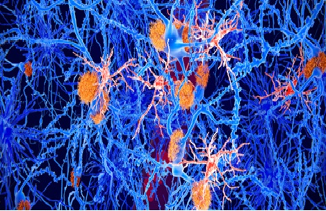
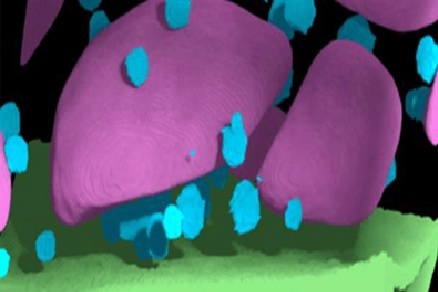
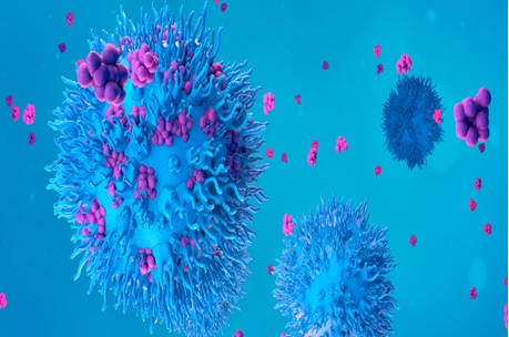
Figure .1 A New Organoid Model Mimics the Growth of Microglia
Figure 1 shows Researchers have developed a novel approach to study the development and behaviour of microglia, the immune cells of the brain, using living human brain tissue models. They colonized cortical organoids, created from human stem cells, with erythromyeloid progenitors (EMPs), which are precursors of microglia. These colonized organoids were then engrafted into immunocompromised mice, leading to vascularization and maturation of the EMPs into functional human myeloid cells within the organoid. Importantly, these cells did not spread to the host.
Using in vivo two-photon microscopy, the researchers were able to observe and monitor the behaviour of the microglia in response to changing environmental factors, such as inflammation and local injuries. This innovative model provides a unique opportunity to study the development and dynamics of microglia in a living human brain tissue context, offering valuable insights into their functions and responses in various conditions. By utilizing fluorescence-activated cell sorting and single-cell RNA sequencing, the research team identified the protein SALL1 as a key regulator of microglial identity and function in their living human brain tissue model. Additionally, they identified specific proteins necessary for the proper functioning of microglia.
Building upon previous research on macrocephalic autism spectrum disorder, which showed abnormal neuronal growth and connectivity, the team investigated the impact of these neuronal differences on microglial development. They compared microglia derived from individuals with macrocephalic autism spectrum disorder to those from neurotypical individuals with macrocephaly. The study revealed that the observed neuronal differences persisted in the model and resulted in microglia that were more responsive to injury or infection. This finding potentially explains the increased inflammation often observed in the brains of individuals with autism spectrum disorder. These findings provide valuable insights into the interplay between neuronal and microglial abnormalities in autism spectrum disorder.
While the results of the study are intriguing, it's important to note that the sample sizes were small, with only three individuals in each cohort. Therefore, the findings should be interpreted with caution, and further investigations with larger sample sizes are necessary to draw more conclusive insights. The research team acknowledges the need for larger studies to validate and expand upon their proof-of-concept findings. They are eager to conduct further investigations using this methodology to study other developmental and neurodegenerative diseases.
Simon Schafer, the first author of the study from the Technical University of Munich, Germany, expressed the team's enthusiasm for applying their brain model to explore the intricate relationship between the brain and immune system. By developing their own brain model, they hope to gain a deeper understanding of these complex interactions and uncover insights that may not be apparent through traditional approaches. The team is committed to refining their model and advancing our understanding of the brain-immune system relationship in the future.
References:
- https://www.biotechniques.com/cell-and-tissue-biology/new-organoid-model-mirrors-microglia-development/
Cite this article:
Janani R (2023),A New Organoid Model Mimics the Growth of Microglia, AnaTechMaz, pp.195
Recent Post
-
CRISPR Gene Editing to The Next Level: No Viruses Needed
The gene-editing technology CRISPR/Cas9, which has revolutionised biomedical research, is well known......
-
One Cell Can Be Recognised and Captured by A Tiny Hybrid Robot
A micro-robot the size of a single biological cell that can recognise and capture a single cell has been.......
-
Possible Connection Between Mom's Vitamin D Intake and Schizophrenia Origins
Researchers have made a significant discovery regarding the role of .....
-
The First Published Draught of The Human "Pangenome" Captures DNA Diversity
The initial version of the human "pangenome," which depicts....
-
A Grant Has Been Given to Develop Volume Electron Microscopy Teaching Materials
The creation of instructional resources for volume electron microscopy.....
-
Human-Machine Interaction Is Being Muddled: Tissue Electrode Growth
A recent study raised the bar for "biotechnology" significantly.....
-
Making Every Effort in The Mab Manufacturing Process
The biopharma industry is making significant strides in antibody development, but it also...
-
Mapping The Connectome of The Fruit Fly Larva
The sites of 3,016 neurons and their connections have been traced in detail for the first time in a fruit fly....
-
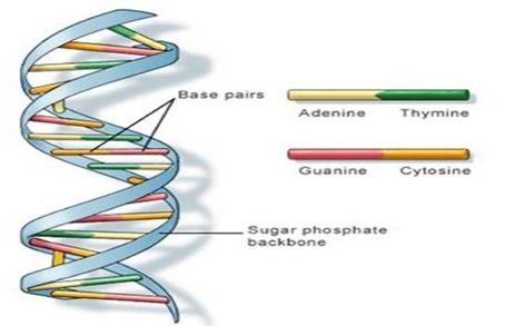
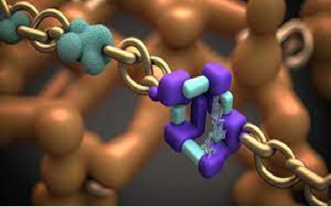
Nanotechnology methods for DNA isolation
Deoxyribonucleic acid (DNA) is the self-replicating genetic material that exists in nearly all living organisms......
-
The Scale-Up Problem in Bioprocessing May Be Solved by Microbioreactors
Volkert van Steijn, an associate professor of chemical engineering ....
-

Risk Management in Biotechnology Innovation
In order to break down industry barriers and decrease failure rates, biotechnology companies ......
-
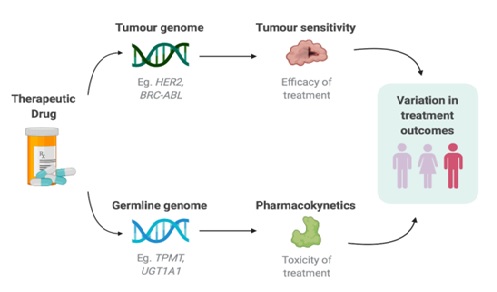
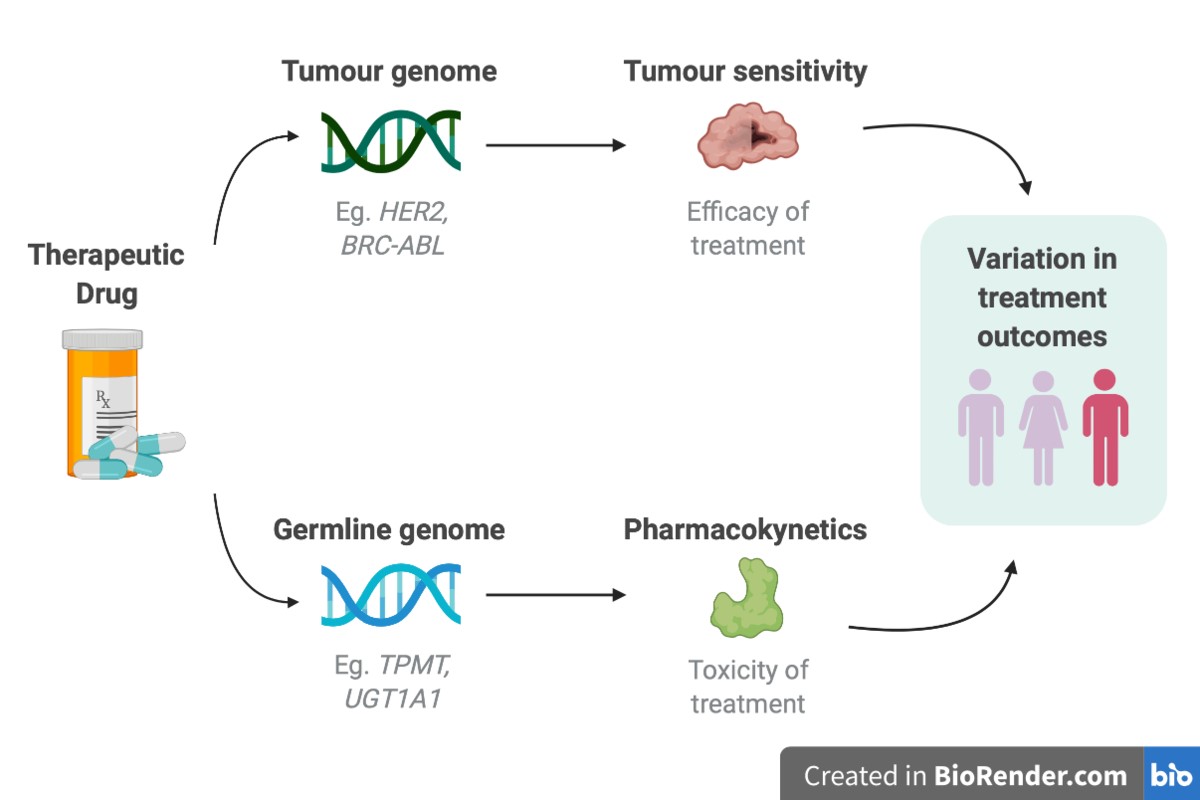
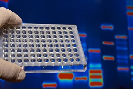
Pharmacogenomics of anti-cancer Drugs
Pharmacogenomics is based on the understanding of the individual differences in drug use, the .....
-
Unveiling the Molecular "Superpower" of Antibiotic-Resistant Bacteria
A recent study conducted by Lund University in Sweden has shed .......
-
Digest Of Technology Ready, Set, Flow: Advancing CAR-T Treatment with Flow Cytometry
The eBook discusses the challenges faced in developing and .....
-
A New Organoid Model Mimics the Growth of Microglia
Fascinating new insights into the growth and operation of microglia have been made possible by a ....