
A Grant Has Been Given to Develop Volume Electron Microscopy Teaching Materials
The creation of instructional resources for volume electron microscopy has received funding
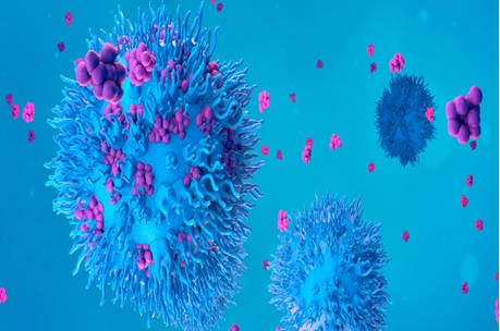
Electron microscopy is a powerful imaging technique, but it is limited to capturing surface images of samples. However, volume electron microscopy (vEM) is an advanced technique that enables scientists to obtain 3D images of larger samples, measuring many cubic millimeters. This breakthrough overcomes the previous need to stack multiple 2D images to achieve a 3D representation. Recognized as one of Nature's technologies to watch in 2023, vEM holds great promise for capturing detailed volumetric information in various scientific fields.
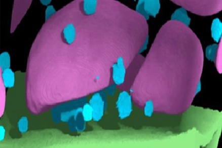
Figure .1 A Grant Has Been Given to Develop Volume Electron Microscopy Teaching Materials
Figure 1 shows The Chan Zuckerberg Initiative has awarded a 2-year grant to an international team for advancing imaging through collaborative projects. Kirk Czymmek and his team at the Donald Danforth Plant Science Center's Advanced Bioimaging Laboratory will utilize the grant to acquire a new volume electron microscope called Helios 5 "Hydra" DualBeam. This cutting-edge microscope will enable them to explore cells and tissues at a cellular level, providing a novel perspective for their research. The grant signifies an exciting opportunity to advance imaging capabilities and enhance understanding in the field of bioimaging.
Kirk Czymmek, director at the Donald Danforth Plant Science Center's Advanced Bioimaging Laboratory, expressed excitement about the potential of the newly acquired Helios 5 "Hydra" DualBeam volume electron microscope. Czymmek highlighted the importance of understanding and improving crop resilience to environmental stress and disease. The advanced capabilities of the microscope will enable the team to reconstruct plant cells and tissues in exceptional detail, creating intricate 3D models that can aid in addressing critical food security challenges. This cutting-edge technology holds great promise for advancing research in the field of plant science and contributing to solutions for global food security.
Kirk Czymmek aims to address the challenges associated with the limited availability of volume electron microscopes (vEM) and the lack of standardized software and training material. To remove these barriers, he plans to develop multimedia outreach materials and training videos, create vEM software platforms, plugins, and reference datasets, establish an infrastructure for a network of vEM-trained scientists, and build a global network of vEM systems. These initiatives will facilitate access to vEM technology, enhance training opportunities, and promote collaboration among scientists working with vEM, ultimately advancing the field and maximizing the potential impact of this innovative imaging technique.
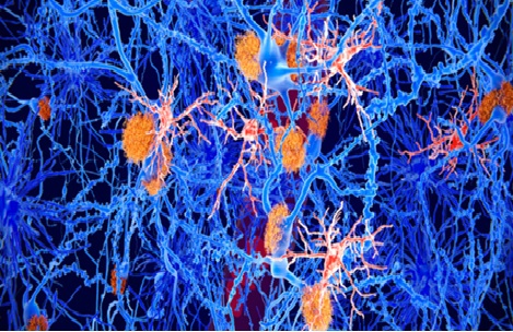
In the coming decades, high-resolution reconstruction of the complete mouse brain may be possible using virtual reality microscopy (vEM), which is already being employed to offer fresh insights into the biology of cancer and infectious illnesses. In the words of Czymmek, "the sooner our colleagues across the globe are trained and educated for successful vEM protocols, the faster new science can be enabled and translate into new medicines and improved foods for the benefit of society."[1]
References:
- https://www.biotechniques.com/lab-design-machinery/10x_sptl_so_grant-awarded-to-create-training-materials-for-volume-electron-microscopy/
Cite this article:
Janani R (2023),A Grant Has Been Given to Develop Volume Electron Microscopy Teaching Materials, AnaTechMaz, pp.185
Recent Post
-

CRISPR Gene Editing to The Next Level: No Viruses Needed
The gene-editing technology CRISPR/Cas9, which has revolutionised biomedical research, is well known......
-
One Cell Can Be Recognised and Captured by A Tiny Hybrid Robot
A micro-robot the size of a single biological cell that can recognise and capture a single cell has been.......
-
Possible Connection Between Mom's Vitamin D Intake and Schizophrenia Origins
Researchers have made a significant discovery regarding the role of .....
-

The First Published Draught of The Human "Pangenome" Captures DNA Diversity
The initial version of the human "pangenome," which depicts....
-
A Grant Has Been Given to Develop Volume Electron Microscopy Teaching Materials
The creation of instructional resources for volume electron microscopy.....
-
Human-Machine Interaction Is Being Muddled: Tissue Electrode Growth
A recent study raised the bar for "biotechnology" significantly.....
-
Making Every Effort in The Mab Manufacturing Process
The biopharma industry is making significant strides in antibody development, but it also...
-
Mapping The Connectome of The Fruit Fly Larva
The sites of 3,016 neurons and their connections have been traced in detail for the first time in a fruit fly....
-

Nanotechnology methods for DNA isolation
Deoxyribonucleic acid (DNA) is the self-replicating genetic material that exists in nearly all living organisms......
-
The Scale-Up Problem in Bioprocessing May Be Solved by Microbioreactors
Volkert van Steijn, an associate professor of chemical engineering ....
-

Risk Management in Biotechnology Innovation
In order to break down industry barriers and decrease failure rates, biotechnology companies ......
-
Pharmacogenomics of anti-cancer Drugs
Pharmacogenomics is based on the understanding of the individual differences in drug use, the .....
-

Unveiling the Molecular "Superpower" of Antibiotic-Resistant Bacteria
A recent study conducted by Lund University in Sweden has shed .......
-
Digest Of Technology Ready, Set, Flow: Advancing CAR-T Treatment with Flow Cytometry
The eBook discusses the challenges faced in developing and .....
-
A New Organoid Model Mimics the Growth of Microglia
Fascinating new insights into the growth and operation of microglia have been made possible by a ....