
Development of Optical Imaging
Optical imaging significantly reduces patient exposure to harmful radiation by using non-ionizing radiation, which includes visible, ultraviolet, and infrared light. Because it is much safer than [1] techniques that require ionizing radiation, like x-rays, optical imaging can be used for repeated procedures to monitor the progression of figure 1 shown below disease or the results of treatment.

Figure1: Optical Imagine
Optical imaging is particularly useful for measuring multiple properties of soft tissue. Because of the wide variety of ways different soft tissues absorb and scatter light, optical imaging can measure metabolic changes that are early markers of abnormal functioning of organs and tissues.
Novel optical imaging for more accurate glaucoma screening. Glaucoma is a leading cause of blindness. Early diagnosis and treatment can slow or stop progression to blindness, but the current test for elevated intraocular [2] pressure is inadequate for detecting glaucoma at an early stage. Changes in the collagen fibers of the sclera (white of the eye) play a significant role in the disease process, potentially offering a reliable marker of early-stage glaucoma.
Optical fiber is the technology associated with data transmission using light pulses travelling along with a long fiber which is usually made of plastic or glass. Metal wires are preferred for transmission in optical fiber communication as signals travel with fewer damages. Optical fibers are also unaffected by electromagnetic interference. The fiber optical cable uses the application of total internal reflection of light. The fibers are designed such that they facilitate the propagation of light along with the optical fiber depending on the requirement of power and distance of transmission. Single-mode fiber is used for long-distance transmission, while multimode fiber is used for shorter distances. The outer cladding of these fibers needs better protection than metal wires.
Optical Coherence Tomography (OCT) is a technique for obtaining sub-surface images such as diseased tissue just below the skin. Ophthalmologists use OCT to obtain detailed images from within the retina. Cardiologists also use it to help diagnose coronary artery disease.
Photoacoustic Imaging delivers laser pulses to a patient’s tissues; the pulses generate heat, expanding the tissues and enabling their structure to be imaged. The technique can help monitor blood vessel growth in tumors, detect skin melanomas, and track blood oxygenation in tissues.
Diffuse Optical Tomography (DOT) and Imaging (DOI) are non-invasive techniques that use light in the near-infrared region to measure tissue properties such as total hemoglobin concentration and blood oxygen saturation. Because DOT and DOI work well in soft tissue, the techniques are widely used for breast cancer imaging, brain functional imaging, stroke detection, photodynamic therapy, and radiation therapy monitoring.
References:
- https://www.nibib.nih.gov/science-education/science-topics/tissue-engineering-and-regenerative-medicine#:~:text=Regenerative%20medicine%20is%20a%20broad%20field%20that%20includes ,to%20recreate%20cells%20and%20rebuild%20tissues%20and%20organs.
- https://byjus.com/physics/what-is-optical-fiber/
Cite this article:
Nandhinidwaraka.S (2021) Development of Optical Imagine, AnaTechMaz, pp. 22
Recent Post
-
Edwards SAPIEN 3 and SAPIEN 3 Ultra Transcatheter Heart Valve (THV) System
The Edwards SAPIEN 3 and SAPIEN 3 Ultra Transcatheter Heart Valve...
-
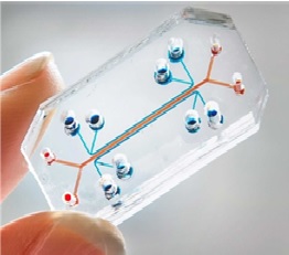
Multiple Uses of Organ-On-a-Chip (OOC)
Although multiple publications claim to have translated organ functions onto this interface, the movement...
-
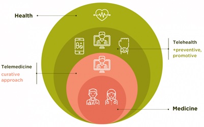
The Difference Between Telehealth and Telemedicine flourish
The healthcare industry is often considered to be the one industry that can work independently...
-
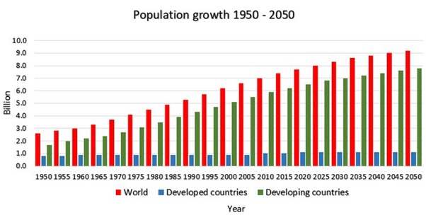
Agriculture in 2050
Transforming agricultural systems and rural economies is the need of the hour if we want to ensure a food-secure future. With the global population...
-

Collaborative BI on the Rise
Collaborative business intelligence (BI) is not an entirely new trend. However, the ever-evolving business landscape, typified by managers...
-
Computer-Supported Cooperative Work (CSCW)
Computer-supported cooperative work (CSCW) consists of software tools and technology that supports a group...
-
Development of Optical Imaging
Optical imaging significantly reduces patient exposure to harmful radiation by using non-ionizing radiation, which includes visible, ultraviolet...
-

Digital Learning, Training, & Development
Training and Development teams have been asked to provide better services and make a bigger impact...
-
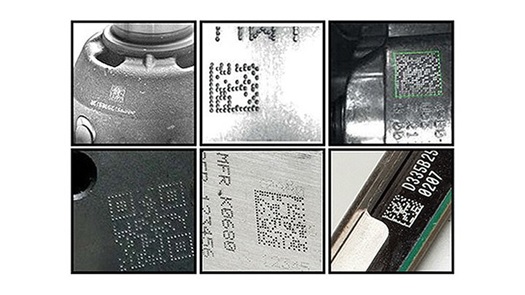
Direct Part Marking (DPM)
Direct Part Marking (DPM) is a technology used to produce two different surface conditions on an item. These markings can be created...
-

Real-Time Locating Systems (RTLS) in Healthcare
Real-time locating systems (RTLS, also known as real-time location systems) have become an important...
-
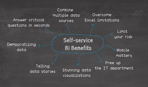
Self-Services BI Interfaces
Self-service BI is a trend with a somewhat vague definition. In the most general sense, self-service BI tasks are those that business users carry...
-

Smart Eye Technology
Smart Eye Technology protects businesses that share highly sensitive files such as financials, legal contracts, wiring instructions, employee...
-
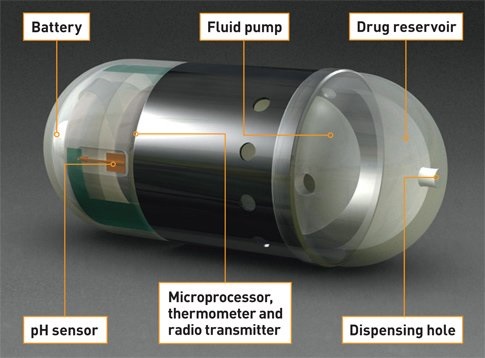
The Features of Micro-Electronic Pills
We are familiar with a wide range of sensors in the field of electronics. They are used widely in the various...
-

The Hyperloop Will Switch Faster Than Aeroplane
Hyperloop is basically a open source project that was first introduced by Elon Musk in 2012.So the basic idea...
-
The Technology of Palm Vein
Palm vein scanning, also known as palm vein recognition, is a modern authentication technology based on biometrics. It utilizes...