
New Imaging Technique to Detect Complications Early in Pregnancy
Oregon Health & Science University researchers have developed a new imaging method to measure the health of a placenta, which could help clinicians identify complications early in a pregnancy.
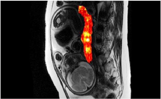
Figure 1: New imaging technique to detect complications.
Figure 1 shows that researchers used magnetic resonance imaging, commonly known as MRIs, and could be replicated on virtually all modern MRI scanners. With quick data analysis, researchers noted that the imaging method could be easily adopted by clinicians. [1]
“Any research that helps us find ways to improve prenatal care is crucial,” said Victoria HJ Roberts. The placenta is a dynamic organ that evolves over the course of pregnancy to support fetal development, so poor placental function early in pregnancy can become an ongoing and increasing health concern both to mother and baby. [2]
Despite the detrimental impact of abnormal placental development, existing methods for evaluating placental function are often ineffective and limited in their ability to reliably detect risks during pregnancy. In prenatal care settings, most clinicians rely on ultrasound to take measurements of fetal growth and blood flow, but this method is limited in scope.
Team to explore how an MRI could be used to give clinicians a more detailed look at placental health than the traditional ultrasound provides, and to better understand an MRI’s effectiveness in detecting placental abnormalities during pregnancy.
The study gathered data from 316 pregnant women across two sites. The OHSU research team developed and validated an MRI protocol that detects a signal in the blood that is linked to oxygen content. This readout is known as T2*, and T2* values provide key information about oxygen availability and placental blood flow.
Oxygen is key for fetal growth and development, so if these values deviate from the normal range, it suggests that something might be wrong. T2* values outside of the normal range could indicate an issue related to the maternal blood supply of oxygen, compromised placental transport or fetal utilization of oxygen.
The study first established a baseline to determine what occurs throughout the course of an uncomplicated pregnancy. Participants underwent three MRI studies during weeks 10 through 40 of pregnancy. Researchers then looked at the ability of MRI to successfully identify complications in pregnancy using the T2* readings produced from the procedures.
The study results suggest that even data from early on in pregnancy — 10 to 20 weeks — can be effective in the identification of at-risk pregnancies. [3]
References:
- https://medicalxpress.com/news/2022-07-imaging-method-complications-early-pregnancy.html
- https://dailynewsera.com/2022/07/28/researchers-develop-new-imaging-method-to-detect-complications-early-in-pregnancy/
- https://news.ohsu.edu/2022/07/27/ohsu-researchers-develop-new-imaging-method-to-detect-complications-early-in-pregnancy
Cite this article:
Sri Vasagi K (2022), New Imaging Technique to Detect Complications Early in Pregnancy, AnaTechMaz, pp.60
Recent Post
-
Cancer Outcomes Predicts by Calculating Tumor Specific Total mRNA Level
Researchers at The University of Texas MD Anderson Cancer Center ......
-
Bacteria with recording function capture gut health status
In a recent study published in Science, researchers from the Department......
-

African Pig Swine Fever Vaccine Developed by Vietnam Researchers
Vietnam has developed an African swine fever vaccine for pigs in partnership......
-

Electric nanomotor made from DNA material
A research team led by the Technical University of Munich (TUM) has succeeded for the first time........
-

Bacteria can live in snake and spider venoms
Bacteria are resourceful little organisms. They can live in some of the weirdest, most inhospitable places......
-

First Organic Bipolar Transistor
OLED displays were a breakthrough after LCD screens, greatly aided by the organic nature of the first’s LEDs. Organic materials........
-

The Development of Biofuels: Energy for Transportation
Unlike other renewable energy sources, biomass can be converted directly into liquid fuels, called "biofuels,......
-

Biomass: A Renewable Energy Resource
Biomass power is electricity generated from renewable organic waste that would otherwise be dumped in ......
-

Biopower: Renewable Electricity Generation Technologies
Biopower technologies convert renewable biomass fuels into heat and electricity........
-

Biologist Found a Way to Understand Microorganism Stress
Biologist Found a Way to Understand Microorganism Stress ......
-

The Human Microbiome
The human microbiome hosts bacteria, microorganisms, and other naturally-occurring flora that can help us and harm us. Diet, stress, drugs......
-
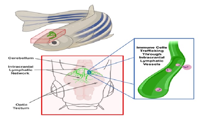
Lymphatics help 'seed' early brain cells in zebrafish
During the embryonic stage of brain development, some neurons and synapses form properly and ......
-

Scientists in US Hack Fruit Flies Brains to Make Them Remote-Controlled
Scientists at Rice University in the United States have figured out........
-

Bacteria Can Remove Plastics from Lakes
Scientists from the University of Cambridgediscovered that some naturally-occurring lake bacteria ........
-
New Imaging Technique to Detect Complications Early in Pregnancy
Oregon Health & Science University researchers have developed a new imaging method......