
Advanced 3D Imaging Uncovers Hidden Skull Changes with Age
Breakthrough 3D Imaging Reveals Aging-Related Changes in Skull's Neurovascular Structures
Researchers from Johns Hopkins University have unveiled the first-ever 3D visualizations of how nerves and blood vessels in the murine calvarium evolve with age. Published in Bone Research, their study employs advanced lightsheet microscopy to map neurovascular architecture from birth to 80 weeks. These findings offer groundbreaking insights into skull aging, highlighting the intricate interactions and gradual decline of nerves and blood vessels over time.
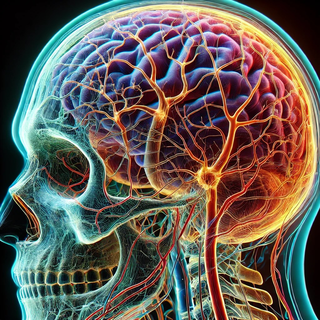
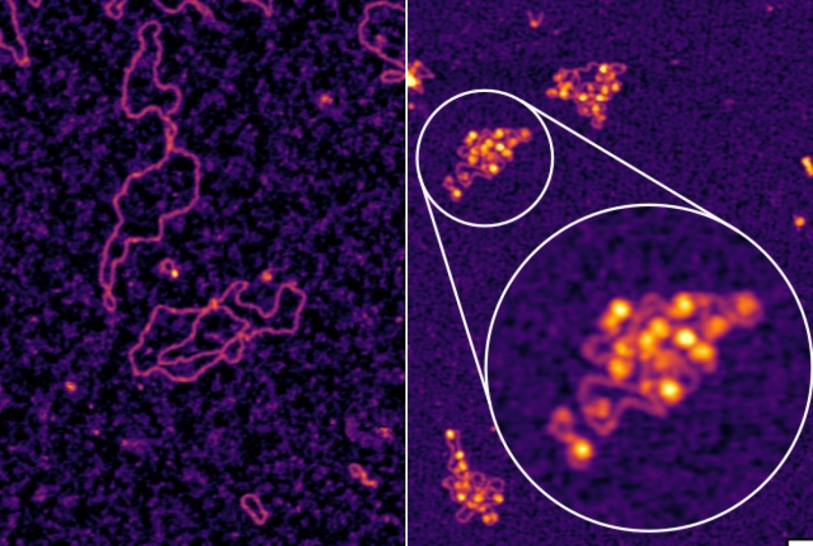
Figure 1. Advanced 3D Imaging Reveals Hidden Changes in the Aging Skull.
3D Imaging Reveals Age-Related Changes in Skull’s Neurovascular Architecture
Researchers from Johns Hopkins University have unveiled the most detailed 3D visualizations of how nerves and blood vessels in the murine calvarium change with age. Published in Bone Research, their study employs advanced lightsheet microscopy to map neurovascular architecture from birth to 80 weeks. Figure 1 shows Advanced 3D Imaging Reveals Hidden Changes in the Aging Skull.
The study revealed a steady increase in nerve density during early life, followed by a significant decline in older mice, particularly in the frontal bone. Blood vessels also exhibited distinct aging patterns, with the relationship between nerves and blood vessels—essential for bone development and regeneration—weakening over time. These changes progressed at different rates across skull regions, with the frontal bone showing the earliest signs of neurovascular decline.
These findings offer groundbreaking insights into the complexity of bone aging, providing valuable data for future research on bone fragility and regenerative medicine.
Implications for Bone Health and Future Therapies
“This research opens up new avenues for understanding how nerves and blood vessels influence bone aging and regeneration,” said Dr. Warren Grayson, one of the lead researchers. “The ability to visualize and quantify these changes in 3D is a significant step forward in our understanding of skeletal health. These insights could help guide future therapeutic strategies for age-related bone diseases and injury recovery.”
By uncovering how neurovascular structures change over time, this study provides a crucial foundation for developing treatments that target bone fragility and enhance regenerative medicine approaches. Understanding these age-related changes may lead to novel interventions aimed at preserving skeletal health and improving recovery from bone injuries.
Transforming Treatments for Bone Diseases and Injury Recovery
The findings of this study have profound implications for treating age-related bone diseases such as osteoporosis and improving recovery from bone injuries. By mapping changes in neurovascular architecture, researchers can better understand the mechanisms underlying bone fragility and impaired healing in older individuals.
These insights could pave the way for innovative therapies that target neurovascular signaling to enhance bone regeneration. Such advancements may lead to more effective treatments for osteoporosis, fractures, and other bone-related conditions, ultimately improving skeletal health and quality of life for aging populations.
Source: SciTECHDaily
Cite this article:
Priyadharshini S (2025),Advanced 3D Imaging Uncovers Hidden Skull Changes with Age,AnaTechMaz,pp. 345
Recent Post
-

Scientists Stunned as Free-Living Coral "Swims" Toward Blue Glow, Breaking the Ruless
Using high-resolution time-lapse imaging, researchers discovered ......
-

Harvard Scientists Uncover Why Humans Burn Energy Differently Than Any Other Animal
Animals consume calories through food, which are then spent on two .

Flagellar Motors: The Secret Behind Bacteria's Near-Perfect Energy Efficiency
Electron microscopy images uncover essential structures and .......

MIT Biologists Uncover a Novel Mechanism for Regulating RNA Splicing
MIT biologists have discovered a new regulatory layer in RNA splicing, revealing a mechanism that influences .....

The type of diabetes influences the risk of heart attack or stroke
A new study has found that individuals with Type 1 diabetes have a lower risk of experiencing cardiovascular .....
-

This Lethal Fungus Manipulates the Immune System, Triggering the Brain to Destroy Itself
A study finds that the fungus Beauveria bassiana alters fruit flies' ....
-
Groundbreaking Discovery Unveils Self-Regulation Mechanism of Key Ion Channel
A recent study conducted by researchers at Weill Cornell ...
-
Tumor "Stickiness" – Researchers Discover a Potential Method to Predict Cancer Spread
“UC San Diego researchers have created a device capable of .....
-
Scientists Uncover Mysterious New Brain Cells That May Revolutionize Alzheimer’s Treatment
Researchers Identify ‘Ovoid Cells’ That Could Unlock New Treatments .....
-
Hepatitis B Enigma Unraveled After Decades – Promising New Treatment Emerges
A team of researchers from Memorial Sloan Kettering Cancer Center ...
-

Scientists Warn: Microplastics Are Silently Fuelling the Rise of Antibiotic-Resistant Superbugs
Microplastics are not just environmental pollutants—they may also .....
-
The Revolutionary Cancer Theory That Could Transform Our Understanding
In a recent essay, scientists challenge the traditional genetic-based...
-
Microscopic Warfare: Bacteria Use Tiny Spearguns for Defense
In the microbial world, peaceful coexistence goes hand in hand with intense competition for nutrients...
-
The Microscopic Struggle That Shapes a Beating Heart
As a heart develops, its cells move, jostling and bumping into each other while searching for their ....
-
Advanced 3D Imaging Uncovers Hidden Skull Changes with Age
Researchers from Johns Hopkins University have unveiled the first-ever 3D visualizations of how nerves ......