
Advancements in 3D Genomic Profiling for Early Pancreatic Cancer Detection
Researchers at the Sol Goldman Pancreatic Cancer Research Center at Johns Hopkins Kimmel Cancer Center have pioneered a 3D genomic profiling technique to identify small precancerous lesions, known as pancreatic intraepithelial neoplasias (PanINs), in the pancreas. These lesions can lead to pancreatic ductal adenocarcinoma (PDAC), one of the most aggressive forms of pancreatic cancer. Their study, published on May 1 in Nature, provides the most detailed 3D map of these precancerous lesions to date, offering a foundation for early detection of PDAC and other pancreatic cancers.
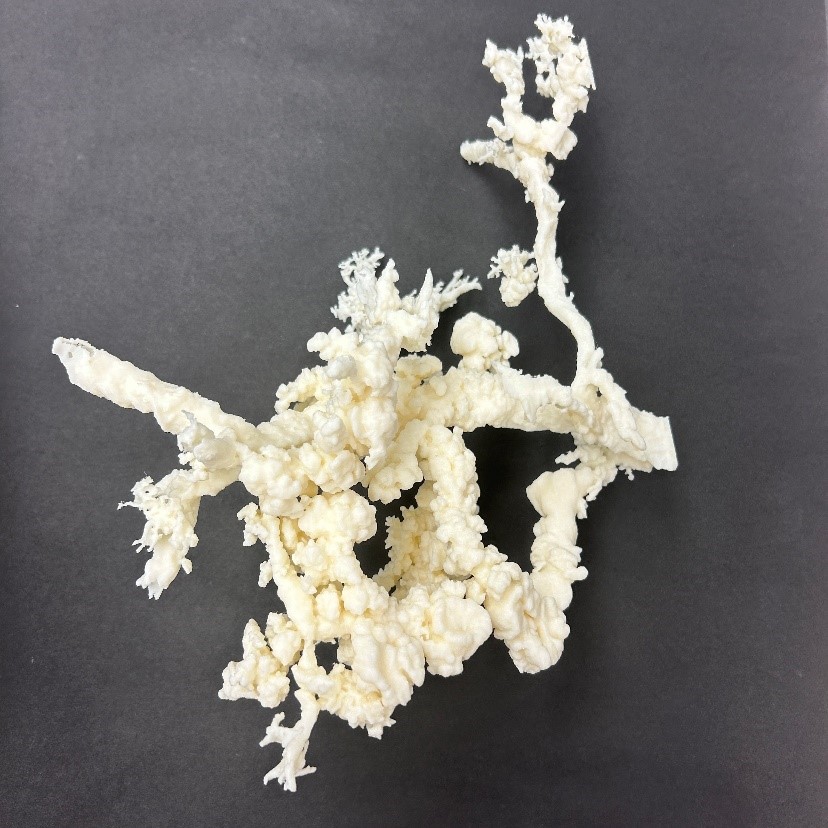
Figure 1. Pancreatic Cancer. (Credit: Ashley Kiemen)
Laura Wood, M.D., Ph.D., associate professor of pathology and oncology at the Kimmel Cancer Center and the Johns Hopkins University School of Medicine, and a senior author of the study, expressed surprise at the high number of PanINs found in normal pancreatic regions. This research highlights the gaps in our understanding of normal aging and the development of cancer in the pancreas. Figure 1 shows pancreatic cancer.
The study was co-led by Alicia Braxton, D.V.M., Ph.D., a postdoctoral fellow, and Ashley Kiemen, Ph.D., an assistant professor of pathology, both from Johns Hopkins University School of Medicine.
PanINs are difficult to detect due to their small size and are not identifiable through typical radiology exams. Consequently, many patients are diagnosed with pancreatic cancer only when it has already reached an advanced and metastatic stage.
Traditional 2D histological staining methods provide a limited view of PanINs, restricting researchers' understanding of their origins and progression to cancer. To overcome this, the researchers developed a 3D approach. They thinly sliced and stained tissue from 38 normal pancreatic samples onto hundreds of sequential 2D slides, then used a machine-learning pipeline called CODA to analyze and reconstruct these images into 3D digital models.
These 3D reconstructions revealed complex networks of interconnected PanINs, averaging 13 PanINs per cubic centimeter, with a range from 1 to 31 PanINs per cubic centimeter. Patients with PDAC in other pancreatic regions had a higher PanIN burden, though this was not statistically significant [1].
Further analysis of eight samples through 3D-guided microdissection and DNA sequencing revealed that these networks comprised genetically distinct PanINs driven by different mutations, including mutations in the cancer-causing gene KRAS, commonly found in pancreatic cancers. This discovery that multiple precancerous lesions can arise from independent mutations is unprecedented in other organs, according to Wood. This finding opens new avenues for targeting these lesions, potentially through KRAS-focused therapies.
Although CODA is not yet ready for clinical diagnostics, Kiemen noted its versatility for application to any tissue, disease, or model organism. She emphasized the importance of continuing research to understand which PanINs are clinically relevant to disease.
Wood concluded that prevention and early detection through detailed 3D molecular maps of cancer precursors could significantly impact cancer treatment. This study marks the beginning of deeper explorations into the early stages of cancer development.
Source: Johns Hopkins Medicine
References:
- https://www.eurekalert.org/news-releases/1048677
Cite this article:
Hana M (2024), Advancements in 3D Genomic Profiling for Early Pancreatic Cancer Detection, AnaTechMaz, pp. 263
Recent Post
-

Unveiling Pathfinder: Advanced Attacks Exploiting Conditional Branch Predictors
Researchers have uncovered two new types of attacks aimed at the conditional branch...
-

Compact, Comfortable, and Immersive Holographic Headset
In the emerging realm of spatial computing, researchers have developed a prototype...
-
Revolutionizing Spinal Injury Treatment: Cambridge's Breakthrough Spinal Cord Device
The development of this tiny, flexible electronic device by a multidisciplinary team at the...
-
Wireless Glaucoma Detection: The Future Potential of 'Smart' Contact Lenses
Early-stage glaucoma often goes undetected, despite the crucial importance of early...
-

Ancient Extinction Mirrors Today’s Ocean Crisis: Deoxygenation’s Role Unveiled
In a groundbreaking discovery, scientists have identified a crucial role of oceanic anoxia in the...
-
Engineers Harness MRI to Detect Deep Brain Light
Researchers often employ the technique of labeling cells with glowing proteins to track tumor...
-

The Impact of Eye Contact on Online Job Interviews: Insights from Recent Research
Eye contact significantly influences interpersonal evaluations, and this extends to online job...
-
Advancements in 3D Genomic Profiling for Early Pancreatic Cancer Detection
Researchers at the Sol Goldman Pancreatic Cancer Research Center at Johns Hopkins...
-
Advancements in Wearable Health Monitoring: Tracking Biochemicals in Sweat
Researchers at Washington State University have developed a wearable health monitor...
-

The Wearable Aptalyzer for Continuous Health Monitoring
Researchers from McMaster University and the University of Waterloo have jointly developed...
-

The Brain's Structure Is Maintained In 'A Fragile Balance.'
When a magnet is heated, it reaches a critical threshold where it loses its magnetization...
-

Human or AI? You Have the Right to Know the Source of What You're Reading
As Google, Microsoft, OpenAI, and now Apple compete to release and improve AI content...
-

The NYPD Has Retired a Large, Egg-Shaped Subway Surveillance Robot—For the Time Being
The New York Police Department's robot, resembling a motionless Wall-E, was seen on...
-

Amazon's Prime Air Delivery Drones Have Received a Significant Clearance from the FAA
The Federal Aviation Administration mandates that commercial drone operators maintain line...
-

Revolutionary Wearable Patch: Real-Time Sweat Monitoring for Optimal Health and Performance
Maintaining bodily water balance is crucial for survival. While sweat serves as an essential...