
DNA Microscopy Reveals 3D Maps of Life at the Molecular Level
Capturing Genes in Action: Imaging Life with DNA
Researchers at the University of Chicago have developed volumetric DNA microscopy, a groundbreaking imaging technique that constructs detailed 3D maps of genetic material. By tagging and tracing molecular interactions, this method reveals unprecedented insights into organisms like zebrafish embryos.

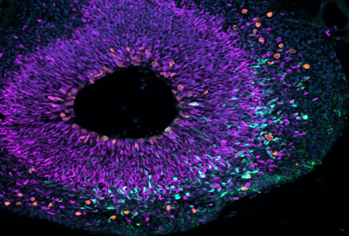
Figure 1. DNA-Based 3D Imaging Unlocks Molecular Insights
A Fresh Perspective on Genetics
Traditional genetic sequencing provides detailed information about the genetic material in a sample, such as tissue or blood, but it lacks spatial context, making it difficult to determine the precise location of specific sequences or their interactions with neighboring genes and molecules. Figure 1 shows DNA-Based 3D Imaging Unlocks Molecular Insights.
To overcome this limitation, University of Chicago researchers are developing volumetric DNA microscopy, a technique that captures both the identity and spatial location of genetic material [1]. By tagging individual DNA or RNA molecules and tracking their interactions, scientists construct a molecular network that maps genetic activity in three dimensions. This approach enables the creation of detailed 3D images of entire organisms at the cellular level, providing unprecedented insights into genetic organization.
Mapping an Entire Organism
Joshua Weinstein, PhD, an Assistant Professor of Medicine and Molecular Engineering at UChicago, has dedicated over a decade to developing DNA microscopy with support from the NIH and NSF. In a study published on March 27 in Nature Biotechnology, Weinstein and postdoctoral researcher Nianchao Qian used the technique to generate a complete 3D DNA map of a zebrafish embryo, a key model for studying development and the nervous system.
“This reveals a level of biology never seen before,” Weinstein said. “Seeing nature from within a specimen in this way is truly exhilarating.”
Redefining Microscopy
Unlike traditional microscopes that rely on light and lenses, DNA microscopy constructs images by analyzing molecular interactions, enabling 3D visualization of genetic material. The process begins by introducing short DNA sequence tags, known as unique molecular identifiers (UMIs), which attach to DNA and RNA molecules. These tags replicate, triggering a chemical reaction that generates unique event identifiers (UEIs), which are specific to each molecular pairing.
These molecular pairings are key to constructing a spatial map of genetic material. UMI pairs in close proximity interact more frequently, producing more UEIs than those farther apart. After sequencing the DNA and RNA, a computational model analyzes these physical connections to reconstruct their original locations, generating a detailed spatial map of gene expression.
Cell Phones and Cells: A Smart Comparison
Weinstein likens the technique to tracking cell phone signals to map people’s locations in a city. Just as knowing a phone’s number or IP address provides identity, tracking its interactions with nearby phones reveals its position. Similarly, DNA microscopy maps molecules by analyzing their biochemical interactions, redefining imaging by using DNA itself instead of light.
Advancing Cancer Research and Immunotherapy
DNA microscopy operates without requiring prior knowledge of a genome or specimen shape, making it valuable for studying genetic expression in unfamiliar contexts [2]. For instance, tumors constantly develop new genetic mutations, and this technique could map their microenvironment and interactions with the immune system. Additionally, since immune cells communicate and respond to pathogens in highly specific ways, DNA microscopy could help decode these genetic mechanisms. Such insights may lead to more precise cancer immunotherapies and personalized vaccines. “This technology bridges a crucial gap in our ability to analyze unique tissue environments,” said Weinstein.
References:
- https://phys.org/news/2025-03-dna-microscope-3d-images.html?deviceType=desktop
- https://scitechdaily.com/dna-microscopy-creates-3d-maps-of-life-from-the-inside-out/
Cite this article:
Janani R (2025), DNA Microscopy Reveals 3D Maps of Life at the Molecular Level, AnaTechMaz, pp. 362
Recent Post
-
Scientists Identify Unique Human-Specific Trigger for Brain Growth
A recent study by researchers at the German Primate Center and the Max Planck Institute reveals that two.....
-
DNA Microscopy Reveals 3D Maps of Life at the Molecular Level
Researchers at the University of Chicago have developed volumetric DNA microscopy, a ......

The Lesser-Known Protein Crucial for Your Hearing
Epithelial cells, which protect our skin and mucous membranes, form a barrier that shields the body from the ....
Supercomputers Reveal Insights into DNA Repair
Researchers use cutting-edge supercomputers to simulate cellular mechanisms involved in DNA .....