
Spectral Flow Cytometry and Imaging: Going Beyond the Scatter Plot
Flow cytometry is a single-cell analysis technique that employs labelled cell structures or biomarkers to distinguish between cell populations. It measures fluorescence intensity of various biomarkers, cell size, and granularity. However, due to overlapping emission spectra, only a limited number of fluorophores can be used simultaneously.
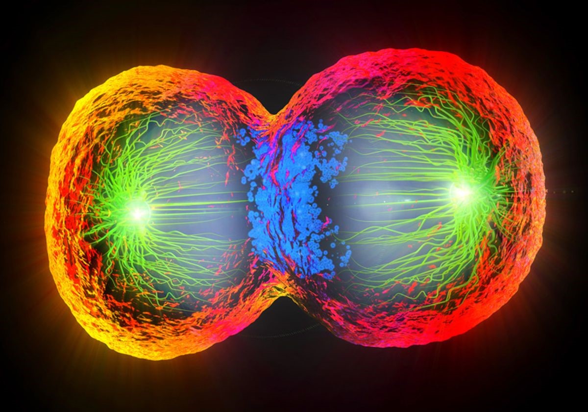
Figure 1. Spectral Flow Cytometry and Imaging: Going Beyond the Scatter Plot
Variations in Flow Cytometry
Figure 1 shows Scientists have developed various adaptations of flow cytometry to enhance its versatility, such as fluorescence-activated cell sorting (FACS), imaging flow cytometry, and spectral flow cytometry. FACS utilizes conventional flow cytometry to identify specific cell populations and enables sorting these cells into separate containers for further analysis or growth. This technique is valuable for isolating distinct cell types or cells with specific protein expressions.
Imaging flow cytometry merges flow cytometry and microscopy, allowing analysis of fluorescently labelled cells' morphology and fluorescence. The approach combines brightfield illumination with fluorescence, capturing both types of images. [1] It offers detailed intracellular and extracellular information, excludes debris and clusters, and provides precise cell size measurements. This method is also high-throughput, enabling faster analyses than traditional microscopy.
Spectral flow cytometry is an advanced version that examines the full emission spectrum of fluorophores, unlike conventional cytometers that assess only limited portions. This capability enables the differentiation of fluorophores with similar emissions, thus expanding the number of simultaneously usable fluorophores for distinguishing cell populations. Although some combinations of these techniques have been employed before, recent advancements have integrated their benefits more comprehensively.
A Creative and Effective Approach
The BD FACSDiscover™ Cell Sorter, developed by BD Biosciences, represents a groundbreaking innovation by integrating FACS, imaging flow cytometry, and spectral flow cytometry into a single instrument. This pioneering technology combines real-time imaging for morphology analysis with flow cytometry data to enable precise identification and sorting of distinct cell subgroups.
Employing 78 fluorescent detectors, the BD FACSDiscover™ S8 Cell Sorter fully captures the emission spectrum of fluorophores, allowing simultaneous use of a wide range of fluorophores. The instrument facilitates rapid sample sorting, capable of isolating six specific cell populations into 5mL collection tubes.[2] The incorporation of real-time images enhances sorting decisions by providing spatial information about both fluorophores and cells.
The BD FACSDiscover™ S8 Cell Sorter employs BD Cell View™ Image Technology, utilizing electronic and optical components for camera-free cell imaging, resulting in faster imaging speed. The system generates multicolour fluorescent and brightfield-like images. Beyond traditional scatter and fluorescence analysis, it empowers researchers to assess 12 distinct cell image features, including parameters like correlation, eccentricity, delta centre of mass, and diffusivity.
With the added advantage of being adaptable to custom biosafety cabinets, the BD FACSDiscover™ S8 Cell Sorter serves a broad spectrum of research fields, including cell biology, oncology, microbiology, and immunology. It enables investigations into diverse areas such as phagocytosis, tumour cell behaviour, and the cell cycle. In summary, the BD FACSDiscover™ Cell Sorter, incorporating BD Cell View™ Image Technology and SpectralFiX™ Technology, amalgamates the strengths of multiple flow cytometry techniques, yielding a versatile and revolutionary research tool.
References:
- https://pubs.rsc.org/en/content/articlelanding/2016/lc/c6lc01063f
- https://www.bdbiosciences.com/en-us/products/instruments/flow-cytometers/research-cell-sorters/bd-facsdiscover-s8
Cite this article:
Janani R (2023), Spectral Flow Cytometry and Imaging: Going Beyond the Scatter Plot,Anatechmaz, pp.478