
Robot Make Surgeries with the Help of Meta’s AI Platform
Scientists at the Francis Crick Institute have developed an imaging technique to capture information about the structure and function of brain tissue at subcellular level – a few billionths of a meter, while also capturing information about the surrounding environment.
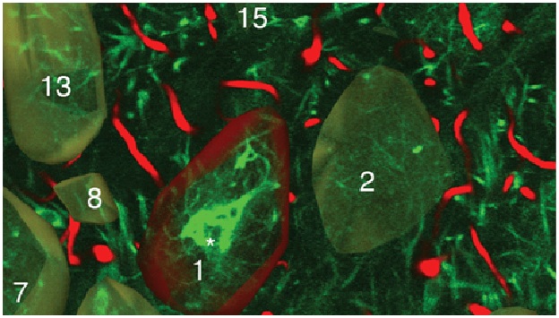
Figure 1: Subcellular level of the brain tissues.
Figure 1 shows that various imaging methods are used to capture information about tissue, cells, and subcellular structures. However, a single method can only capture information about either the structure or function of the tissue and looking in detail at a nanometer scale. This means that to gain an overall understanding of the tissue, imaging techniques need to be combined.
In their study, the scientists developed an approach that combines seven imaging methods, including in vivo imaging, synchrotron X-ray, and volume electron microscopy. They demonstrated their approach by imaging two different areas of the brain in mice – the olfactory bulb and the hippocampus. [1]
Each step of the imaging process provides different information. Firstly, the researchers used in vivo calcium imaging to visualize neurons in specific regions of the brain and see which neurons were active when the mice were exposed to odors.
After the mice were euthanized, brain tissue samples were imaged using various methods, which captures samples up to several millimeters in length. This scale is sufficient for scientists to see whole neural networks and also where particular cells or other structures sit within the wider context of the sample. Importantly, this method does not damage the sample so it can be imaged again using another technique.
In some target areas, this could map details as small as 10 nanometers, allowing researchers to see tiny structures like the individual synapses that connect neurons.
Using computer algorithms, they combined the results to create a complete map of the structure and function of the section of the brain they were studying, up to a few cubic millimeters. [2]
“Our approach provides a reliable way to overcome the challenge of imaging structures at vastly different scales. We believe it will be a powerful tool to investigate neuronal circuits in the brains of mammals as well as the structure and function of other tissues,” says Carles Bosch.
But it also has great potential to be useful in other settings, like cancer biology where researchers aim to understand the activity of particular cells in the context of the wider tumour.
The team will continue this research, using this imaging approach to uncover more detail about the olfactory bulb as well as working to further improve the technique.
References:
- https://technoblender.com/new-imaging-technique-generates-incredible-subcellular-maps-of-entire-brain-networks/
- https://theprint.in/science/scientists-develop-subcellular-map-of-complete-brain-network/970410/
- https://scitechdaily.com/new-imaging-technique-generates-incredible-subcellular-maps-of-entire-brain-networks/
Cite this article:
Sri Vasagi K (2022), Scientist Found Imaging Technique to Capture Brain at Subcellular Level, Anatechmaz, pp. 329