Neuroscientists Develop a Detailed Map of the Cerebral Cortex
Using fMRI, MIT researchers have created the most detailed map of the brain’s cerebral cortex to date. By analyzing brain scans of individuals watching movie clips, they identified 24 distinct networks responsible for various functions such as language, social interactions, visual processing, and sensory input.
While these networks have been observed in previous studies, they have not been fully characterized under naturalistic conditions. Previous research often relied on specific tasks or brain scans taken while subjects were at rest. This new approach, studying the brain during more dynamic scenarios, offers fresh insights into how these networks function.

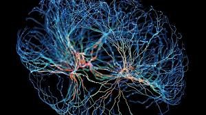
Figure 1. Neuroscientists Map the Cerebral Cortex
“There’s a growing trend in neuroscience to study brain networks in more naturalistic settings. This approach uncovers new perspectives that traditional neuroimaging might miss,” says Robert Desimone, director of MIT’s McGovern Institute for Brain Research. Figure 1 shows Neuroscientists Map the Cerebral Cortex.
This map serves as a foundation for future research, exploring the roles and interactions of these networks. The study, published in Neuron, is co-authored by Desimone and John Duncan from the MRC Cognition and Brain Sciences Unit at Cambridge University. The lead author is Reza Rajimehr, a research scientist at the McGovern Institute.
Detailed Mapping
The cerebral cortex houses regions dedicated to processing various sensory inputs, such as visual and auditory information. Over recent decades, scientists have mapped numerous networks involved in these processes, often using fMRI while subjects perform specific tasks like viewing faces.
In other studies, brain scans are taken while individuals are at rest or allow their minds to wander, which led to the identification of networks such as the default mode network—active during introspective activities like daydreaming.
"Most prior studies on brain networks focused on resting-state fMRI, identifying key networks in the cortex linked to specific cognitive functions. These findings have been central to neuroimaging research," says Rajimehr.
However, during the resting state, much of the cortex may remain inactive. To capture a more complete view of the brain's activity, the MIT team analyzed data gathered as participants engaged in a more natural task—watching a movie.
“Using a rich stimulus like a movie allows us to engage many regions of the cortex more efficiently. Sensory areas activate to process visual and auditory details, while higher-level regions process semantic and contextual information,” explains Rajimehr. “This approach enables us to differentiate networks based on their distinct activation patterns.”
Cognitive Control Networks
Three of the networks identified in this study are associated with "executive control" and were most active during transitions between movie clips. The researchers also observed a "push-pull" dynamic between these control networks and networks that process specific features like faces or actions. When domain-specific networks were highly active, the executive control networks were mostly inactive, and vice versa.
“High activity in domain-specific areas seems to reduce the need for engaging high-level networks,” Rajimehr explains. “However, when the stimulus becomes more complex or ambiguous, the executive control networks become more active.”
By using a movie-watching paradigm, the researchers are further investigating these networks to identify subregions linked to specific tasks. For instance, within the social processing network, they have pinpointed regions involved in recognizing faces and bodies. They’ve also identified areas responsible for processing memories of places within a newly discovered network focused on visual scenes.
“This experiment is aimed at generating hypotheses about how the cerebral cortex is functionally organized. The networks that emerge during movie-watching will now serve as the basis for more specific studies to test these hypotheses, offering us new insights into how the cortex operates during a more naturalistic task compared to resting conditions,” says Desimone.
The study was funded by the McGovern Institute, the Cognitive Science and Technology Council of Iran, the MRC Cognition and Brain Sciences Unit at the University of Cambridge, and a Cambridge Trust scholarship.
Source: MIT News
Cite this article:
Janani R (2024), Neuroscientists Develop a Detailed Map of the Cerebral Cortex , AnaTechmaz, pp.1044

